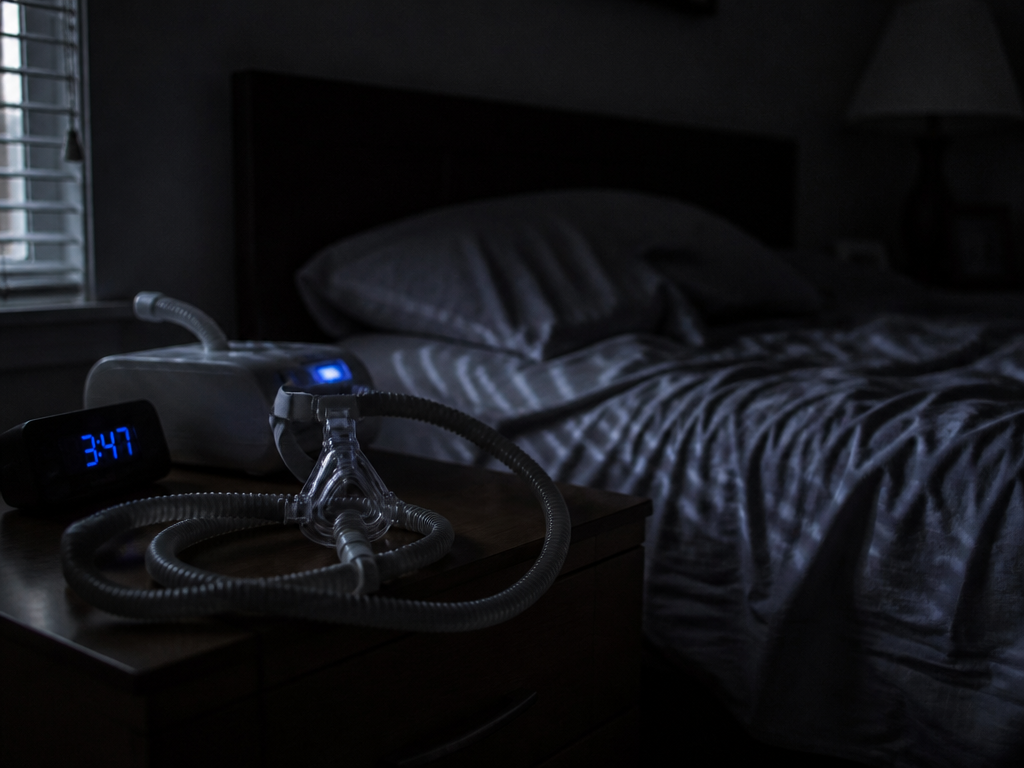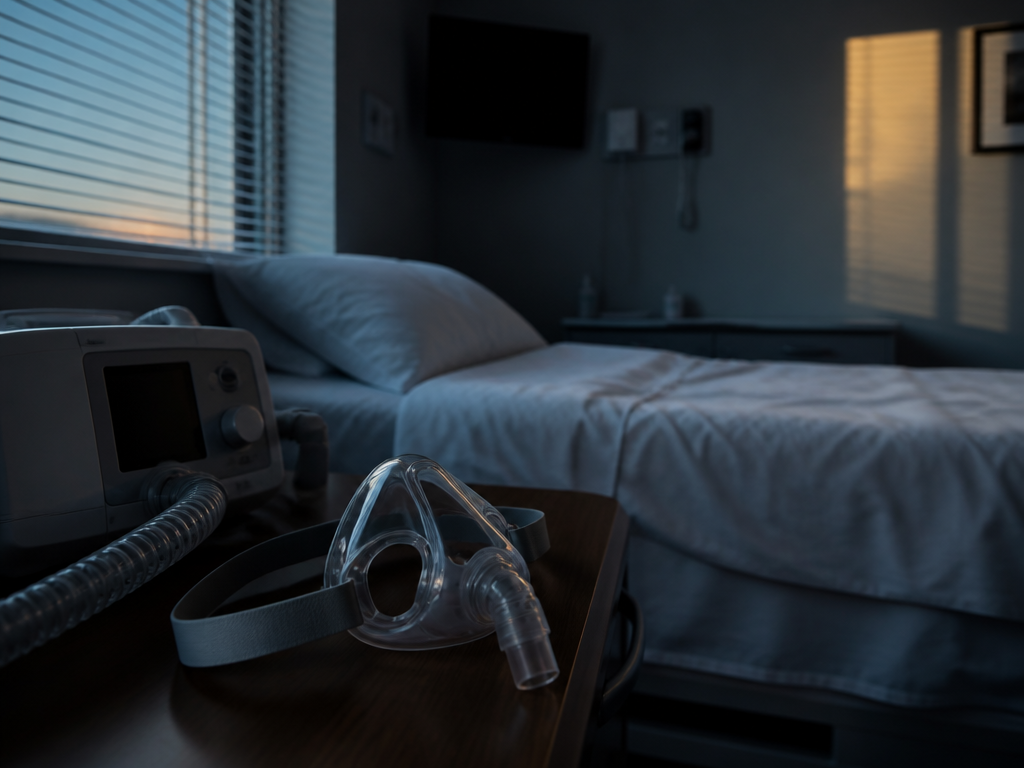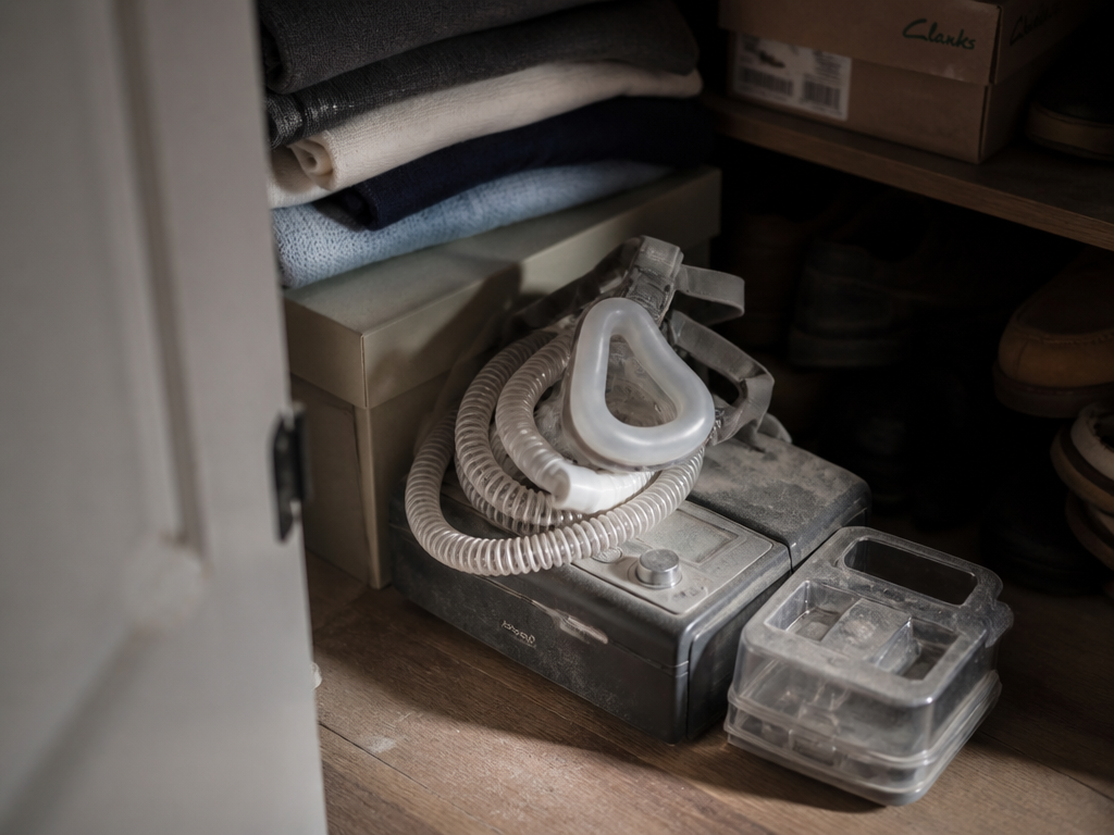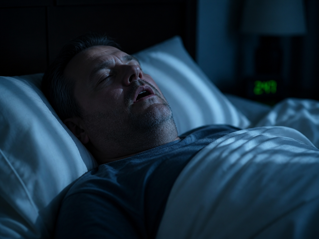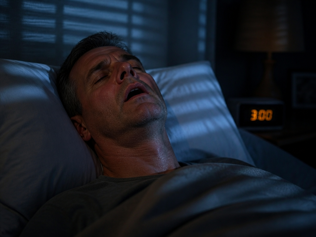
When the upper airway collapses during sleep, the most visible consequence is the one you can hear: the snore that stops, the silence, the gasp. But the quieter consequence happens in the chest. In a meaningful fraction of people with sleep apnea, each obstructed breath is accompanied by a measurable drop in heart rate — bradycardia — that resolves the moment air moves again. The verdict, stated plainly: airway obstruction is not only a respiratory event but a cardiac one, and on overnight data the two move together closely enough that you can often watch one cause the other.
I want to separate three things that get blended in most conversations about this: what people assume is happening, what the published physiology actually shows, and what I observed when I recorded my own nights.
What most people do
Most people — patients and a fair number of well-meaning caregivers — treat obstructive sleep apnea as a pure breathing problem. The mental model is simple: the throat closes, oxygen drops, the brain panics, you wake. The number everyone fixates on is the AHI, the apnea-hypopnea index, which counts how many times per hour breathing stops or shrinks. Under 5 is normal; 15 to 30 is moderate; above 30 is severe. That index is genuinely useful, and I am not dismissing it.
But it describes the airway, not the heart. A patient handed an AHI of 22 rarely asks what their pulse was doing during those 22 events, and the summary report rarely volunteers it. Consumer sleep trackers compound the gap. They graph an average overnight heart rate — a single soft number — which smooths away exactly the thing that matters: the sharp, brief dips that ride on each obstruction. Averaging is the enemy of seeing bradycardia. You can have an alarming beat-to-beat pattern and a perfectly boring nightly average.
So the default behavior is to manage the breathing and assume the cardiovascular system will follow. Sometimes it does. But the mechanism deserves a closer look, because it explains why some apnea patients feel their hearts "skip" at night and why infant apnea is a categorically different worry.
What the evidence suggests
The link runs through the vagus nerve. During an obstructed breath you keep trying to inhale against a closed airway. That effort, combined with falling blood oxygen, triggers a reflex with deep evolutionary roots — the same one seen in diving mammals — in which the body conserves oxygen by slowing the heart. Vagal tone rises, the sinus node fires more slowly, and the rate falls. When the airway finally opens and oxygen returns, sympathetic activity surges and the rate rebounds, often overshooting. The result is a sawtooth: slow during the event, fast on release.
In adults, the literature describes this most often as cyclical variation in heart rate, and in more severe cases as frank nocturnal bradycardia or pauses. The pattern tends to track event severity and the depth of oxygen desaturation — the lower the oxygen dips, the more pronounced the slowing. Importantly, the arrhythmias are usually nocturnal and event-bound: they appear with the obstructions and largely disappear when the obstructions are treated. That last point is the hopeful part of an otherwise serious picture.
Infant apnea is not a smaller version of this. In infants, especially those born preterm, apnea and the accompanying bradycardia more often reflect an immature central drive to breathe rather than a collapsing airway. The bradycardia there is a marker of instability in the brainstem's respiratory control, not a vagal response to mechanical obstruction. The two share a vocabulary and almost nothing else.
| Feature | Adult OSA-related bradycardia | Infant apnea-related bradycardia |
|---|---|---|
| Usual driver | Airway obstruction + vagal reflex | Immature respiratory control |
| When it appears | During obstructive events, in sleep | During apnea spells, often awake or asleep |
| Typical fix | Treat the obstruction (e.g., CPAP) | Maturation, monitoring, sometimes caffeine therapy |
| Reversibility | Often resolves with airway treatment | Usually resolves as the infant matures |
The takeaway is not that every apnea patient has a heart problem. It is that the same word points at two different mechanisms, and conflating them — as headlines often do — leads caregivers to either over-worry or mis-direct their worry.
What I actually do
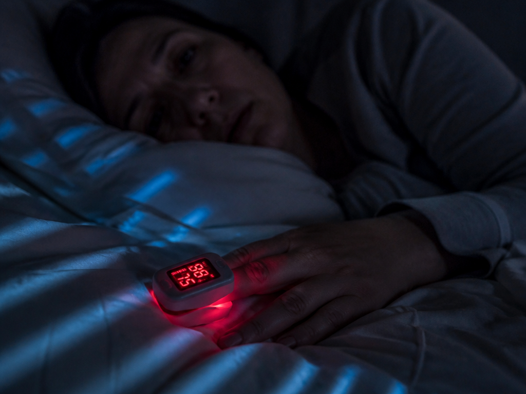
I am one data point with moderate, positionally-driven snoring, not diagnosed severe apnea, so read this as an experiment and not a study. For five nights I wore a recording pulse oximeter that logs SpO₂ and heart rate every few seconds, and I reviewed the traces in the morning rather than trusting a summary score.
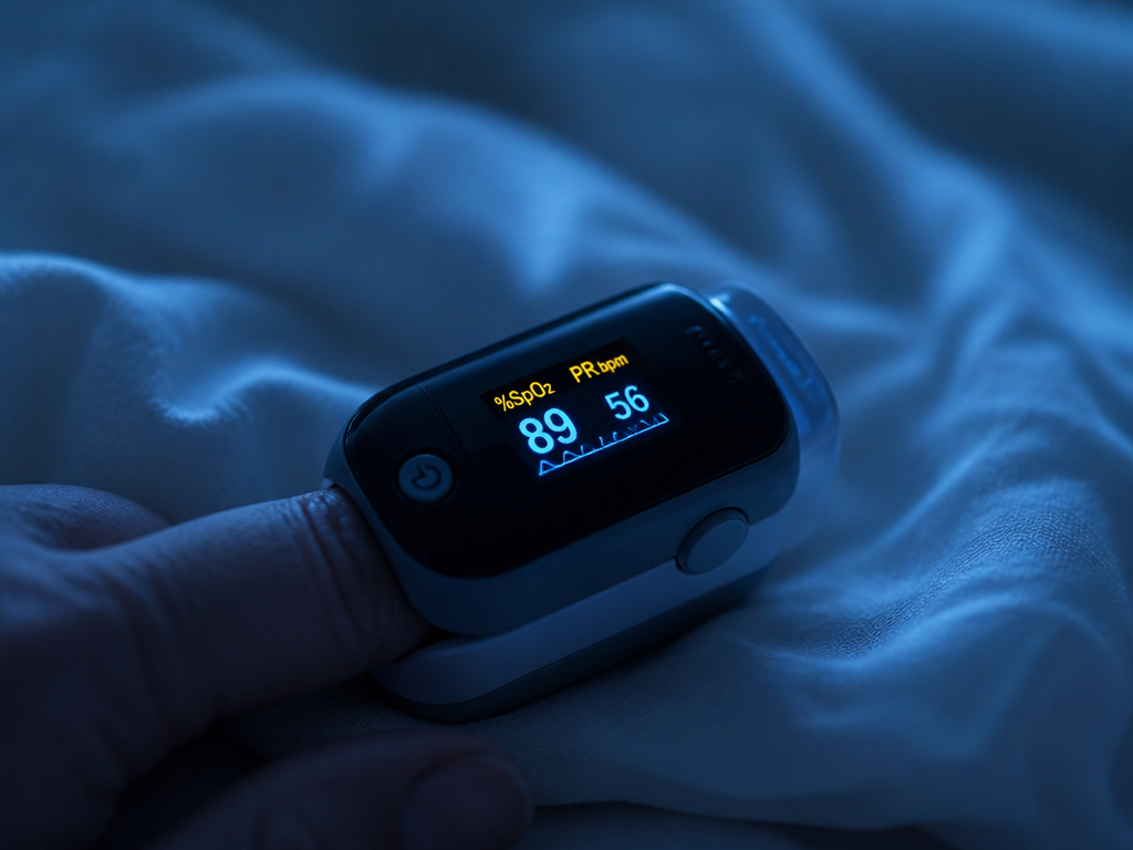
On two of the five nights — both nights I slept on my back — I could see it clearly. Oxygen would slide from 96% down to the high 80s, and trailing that dip by a few seconds the heart rate would fall, in one case from a sleeping baseline near 54 down to 41, then rebound into the 70s within a breath or two of recovery. The dips clustered in the early-morning hours when REM is denser. Side-sleeping nights showed almost none of it.
What I could not test matters. A wrist or finger oximeter is not an ECG; it cannot distinguish a true sinus pause from a motion artifact, and it tells you nothing about the type of rhythm. Five nights is not a trend. And I had no formal sleep study to confirm the events were obstructive rather than central. So my honest conclusion is narrow: in my own data, back-sleeping produced oxygen dips, and heart-rate dips reliably followed them. That is consistent with the published mechanism. It is not a diagnosis, and I'd treat any persistent low-40s reading as a reason to get a real recording, not a home gadget.
Who this is for — and who it isn't
This is for the apnea patient who wants to understand why a partner says their breathing and their pulse seem linked, and for the caregiver who has heard "bradycardia" attached to both a grandparent's apnea and a newborn's monitor and needs to know those are different events. It is not for someone using a smartwatch average to rule out a heart problem; that tool cannot do that job. And it is not a substitute for a sleep study if your daytime sleepiness or witnessed pauses suggest one.
If there is a single line worth keeping: treat the airway and the event-bound heart-rate dips usually go with it.
One thing to try this week
Look at the shape of your data, not the average. If your tracker lets you see a per-minute or beat-to-beat heart-rate graph from a single night, open one full night and scan for sawtooth dips clustered in the pre-dawn hours. Note whether they line up with the nights you slept on your back. You won't diagnose anything — but you'll know whether this conversation is about your body at all, which is more than an average will ever tell you.